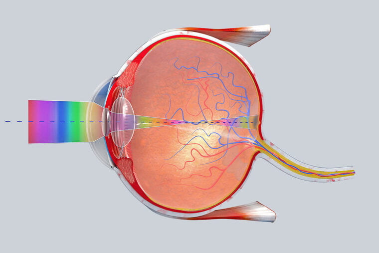
To see how we see the world and how illnesses, for example, age-related macular degeneration and glaucoma disable vision, researchers need to see how the retina imparts vision signs to the brain.
Beforehand, scientists have worked basically with retinal cells from creatures. Be that as it may, another investigation from researchers at Washington University School of Medicine in St. Louis subtleties the analysts’ work with cells from human retinas and how those phones convert visual signs conveyed by the optic nerve to the brain, which at that point deciphers the signs into sight.
In the study, distributed June 12 in the journal Neuron, the specialists report that particular retinal ganglion cells that convey by far most of visual signs from the human retina to the brain productively procedure and pack that data so it tends to be moved.
The researchers took in this by checking the action of human retinal cells while retinas were being presented to films that made those phones transmit flags as the view changed.
“If we want to learn how to correct vision deficits in people who have retinal diseases, as well as problems like glaucoma that damage the optic nerve, we need to know how these ganglion cells convert and transmit signals,” said head specialist Daniel Kerschensteiner, MD. “Visual systems are adapted to specific environments and behavioral demands, and these ganglion cells have adapted to those needs over millions of years of evolution. Although we share certain aspects of our visual systems with animals, if we want to really understand and restore vision in people, we need to better understand how human ganglion cells encode and relay visual information.”
Kerschensteiner, a teacher of ophthalmology in the John F. Hardesty, MD, Department of Ophthalmology and Visual Sciences, and an educator of neuroscience and of biomedical building, drove the investigation of retinal cells from natural eyes that must be expelled because of visual malignant growth.
In spite of the fact that the researchers utilized retinas from just four natural eyes, the specialists had the option to make determinations by considering the action of several ganglion cells in each eye. The axons of those cells consolidate to shape the optic nerve.
The scientists concentrated on the two primary cell types that transmit visual data, every one of which comes in two varieties—one that reacts when light increments and one that reacts when light declines.
Kerschensteiner’s group performed PC investigation of the cells’ movement and discovered that for each situation, these cells transmitted the most extreme measure of visual data utilizing the base measure of vitality.
By utilizing PC displaying to change how the cells carried on, the researchers had the option to show that the ganglion cells were productive. Be that as it may, at whatever point they made those alterations on the PC, signals from the phones turned out to be less effective.
“The optic nerve is a real bottleneck in the visual system,” he clarified. “And it appears from our analysis that these cells evolved to compress visual information to get it through this narrow bottleneck. Patients with eye diseases, such as glaucoma, have difficulty doing that, and to correct those deficits, we need to better understand how healthy cells function. We’re just beginning to gain that understanding.”